Microwaves for BioMedicine

Microwaves for biomedical applications, including dual-mode microwave applicators with integrated microwave sensorics as well as biological sensors with integrated microfluidic channels and underlying measurement techniques for microwave-based analysis and manipulation of biological cells.

Team Leader: Martin Schüßler

Microwave sensor with microfluidic channel for determining the dielectric properties of biological liquids.

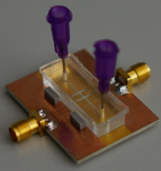
Microwave sensors for biomedical applications provide a non-contact, marker-free, and non-destructive way to monitor processes and systems. The dielectric properties of biological materials in the microwave frequency range from 300 MHz to 300 GHz provide information about the molecular composition of substances. Microwave sensors have a very high flexibility in design and appearance, which makes in-vitro diagnostics and lab-on-chip applications feasible.

Depending on the requirements of the sensor, new geometries and topologies enable to solve the measuring tasks. Our group can fall back on experience of a wide variety of microwave sensor applications in medicine, biology, environmental and industrial process monitoring. Interdisciplinary cooperation always plays a decisive role in order to be able to investigate new and functional materials, innovative processing technologies, microfluidic systems as well as biological processes.

Dual-mode applicator in a two-layer phantom with similar characteristics of a tumor surrounded by liver tissue.

Microwave ablation (MWA) is a promising therapeutic option for the treatment of solid tumors, e.g. in the liver. During the minimally invasive procedure, a needle-shaped antenna is guided to the tumor, which is then destroyed by increasing temperatures. In contrast to surgical resection, critical regions of the body can be reached. A current challenge of MWA is the availability of sufficiently accurate imaging before and during the intervention. Thus, the treatment success still depends on the experience of the radiologist.

We are investigating how microwave technology can be further used for detection before and during the intervention. Thus, the active part of the applicator can be placed exactly in the center of the tumor and treatment monitoring can be realized to minimize the duration of the intervention and to increase the success of the therapy. This work is done in close cooperation with the Institute for Diagnostic and Interventional Radiology at University Hospital in Frankfurt.

Microwave porator in which uptake of peptides has occurred in a field strength-dependent manner. The black dots correspond to the cells in which uptake occurred.

The study of intracellular processes through the targeted imprinting of drugs, DNA, RNA or peptides is necessary to understand the function of biological cells in detail. The cell membrane is a natural barrier between the inside and outside of a cell. Electroporation is one option to bypass the cell membrane that is used frequently in laboratories all over the world. Microwave-induced electroporation is a promising alternative to conventional transfection methods due to the application of lower electric field strengths and a potential higher viability of the cells. We investigate the fundamental functionality of microwave-induced electroporation methods by considering frequency-, time-, and power- dependent relations between a successful uptake of biological substances into cells and their viability. This work is done in a close cooperation with the cell biology and epigenetics lab of the biology department at TU Darmstadt.